%20Wolverine%20Stack%20(20mg).jpg)
BPC-157 / TB-500 Mix
In Stock
Out of Stock
$136
Net Content
10/10mg
Please select a content
{00.00}
{stockCount}
1
Successfully email sent to admin
PASSED
Sterility & Endotoxins
PASSED
Net Content & Purify
Products offered by Pythion Research are provided for research and educational purposes.
Properties
Overview
BPC-157 / TB-500 is a dual-peptide research blend composed of two extensively studied synthetic peptides: BPC-157 (Body Protection Compound-157) and TB-500 (synthetic thymosin beta-4 analog). Each peptide has been investigated in laboratory settings for its potential involvement in cellular repair signaling, angiogenesis pathways, and inflammatory response regulation.
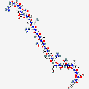
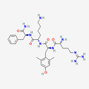
BPC-157 is a 15-amino-acid synthetic peptide fragment derived from a gastric protein sequence, studied in experimental models examining vascular signaling, endothelial protection mechanisms, and tissue regeneration pathways. TB-500 is a 43-amino-acid synthetic analog of thymosin beta-4, a naturally occurring peptide involved in cellular migration and cytoskeletal organization.
Because both peptides influence distinct but interconnected biological systems, researchers have explored their roles in experimental models evaluating tissue remodeling, vascular formation, cellular motility, and inflammatory pathway signaling. Dual-peptide formulations such as this blend allow investigators to examine how complementary peptide mechanisms may interact within complex biological environments.
Research Background
Scientific investigations have examined both BPC-157 and TB-500 across studies related to cellular regeneration, angiogenesis signaling, and extracellular matrix repair.
Research involving TB-500 has explored its interaction with actin-binding proteins and cytoskeletal dynamics, which influence cellular movement and structural organization. Experimental models suggest that TB-500 may regulate actin polymerization by binding to globular actin (G-actin), potentially facilitating cellular migration and angiogenic signaling pathways. Studies have also investigated TB-500’s influence on inflammatory signaling through regulatory molecules such as microRNA-146a, which may affect downstream signaling proteins including IRAK1 and TRAF6.
BPC-157 has been examined in research exploring vascular signaling pathways and endothelial cell stability. Experimental studies suggest that BPC-157 may interact with nitric oxide (NO) signaling mechanisms and pathways involved in cellular adhesion and migration, including FAK-paxillin signaling networks associated with tissue repair processes.
Laboratory investigations have also reported increased collagen formation and vascular development in experimental injury models exposed to BPC-157. These findings have led researchers to examine the peptide in studies focused on connective tissue regeneration and extracellular matrix organization.
Because TB-500 and BPC-157 influence complementary biological mechanisms—including cytoskeletal regulation, vascular signaling, and inflammatory pathway modulation—this peptide combination has been studied in research environments examining complex regenerative signaling systems.
Common Research Focus Areas
Tissue repair and regenerative pathway investigations
Angiogenesis and vascular signaling studies
Cytoskeletal regulation and cellular migration research
Extracellular matrix and collagen formation studies
Inflammatory pathway and immune signaling analysis
Research Peptide Mixture (non-formulated) | BPC-157 (5mg) + TB-500 (5mg) |
Content | 10mg (5mg + 5mg) |
Physical Appearance | Fine White Lyophilized Powder |
Storage | Store in a cool, dry environment. Protect from moisture, heat, and direct light. |
Terms | Products offered by Pythion Research are provided for research and educational purposes. |
2D Structure

Related Products


Sale: 20% Off Sitewide — Use Code: PYTHION20 At Checkout • Ends May 10th